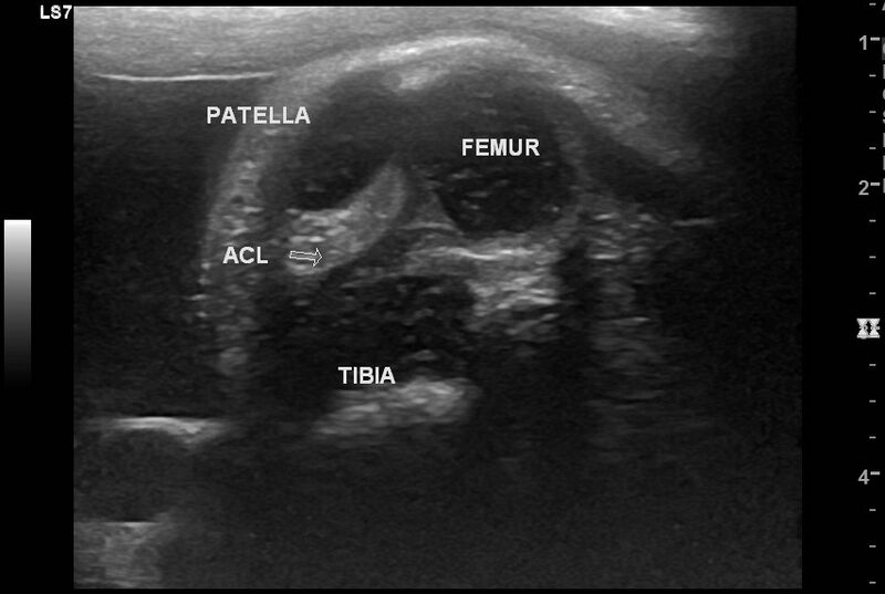
File:Anterior and posterior cruciate ligaments (fetus) (Radiopaedia 82919).jpg
Jump to navigation
Jump to search

Size of this preview: 800 × 537 pixels. Other resolutions: 320 × 215 pixels | 640 × 430 pixels | 853 × 573 pixels.
Original file (853 × 573 pixels, file size: 76 KB, MIME type: image/jpeg)
Summary:
| Description |
|
| Date | Published: 9th Oct 2020 |
| Source | https://radiopaedia.org/cases/anterior-and-posterior-cruciate-ligaments-fetus |
| Author | Maulik S Patel |
| Permission (Permission-reusing-text) |
http://creativecommons.org/licenses/by-nc-sa/3.0/ |
Licensing:
Attribution-NonCommercial-ShareAlike 3.0 Unported (CC BY-NC-SA 3.0)
File history
Click on a date/time to view the file as it appeared at that time.
| Date/Time | Thumbnail | Dimensions | User | Comment | |
|---|---|---|---|---|---|
| current | 14:47, 7 May 2021 | | 853 × 573 (76 KB) | Fæ (talk | contribs) | Radiopaedia project rID:82919 (batch #2103) |
You cannot overwrite this file.
File usage
The following page uses this file:
_(Radiopaedia_82919).jpg&oldid=8854026)