File:Anterior circle of Willis (illustration) (Radiopaedia 36124).jpg
Jump to navigation
Jump to search
Size of this preview: 600 × 600 pixels. Other resolutions: 240 × 240 pixels | 480 × 480 pixels | 640 × 640 pixels.
Original file (640 × 640 pixels, file size: 53 KB, MIME type: image/jpeg)
Summary:
- Radiopaedia case ID: 36124
- Image ID: 14185
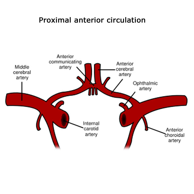
- Study findings: Diagram of the basilar artery and circle of Willis. This diagram has been reproduced from Gray's Anatomy 20th US edition which has now lapsed into the public domain (http://en.wikipedia.org/wiki/Image:Circle_of_Willis_en.svg)
- Modality: Diagram
- System: Vascular
- Findings: Diagram of the basilar artery and circle of Willis. This diagram has been reproduced from Gray's Anatomy 20th US edition which has now lapsed into the public domain (http: //en. wikipedia. org/wiki/Image: Circle_of_Willis_en. svg)
- Published: 15th May 2015
- Source: https://radiopaedia.org/cases/anterior-circle-of-willis-illustration
- Author: Frank Gaillard
- Permission: http://creativecommons.org/licenses/by-nc-sa/3.0/
Licensing:
Attribution-NonCommercial-ShareAlike 3.0 Unported (CC BY-NC-SA 3.0)
File history
Click on a date/time to view the file as it appeared at that time.
| Date/Time | Thumbnail | Dimensions | User | Comment | |
|---|---|---|---|---|---|
| current | 21:43, 18 March 2021 | | 640 × 640 (53 KB) | Fæ (talk | contribs) | Radiopaedia project rID:36124 (batch #2105) |
You cannot overwrite this file.
File usage
The following page uses this file:
_(Radiopaedia_36124).jpg&oldid=8859854)