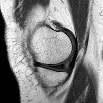
File:Anterior cruciate ligament mucoid degeneration (Radiopaedia 60853-68633 Sagittal T1 38).jpg
Jump to navigation
Jump to search
Anterior_cruciate_ligament_mucoid_degeneration_(Radiopaedia_60853-68633_Sagittal_T1_38).jpg (400 × 400 pixels, file size: 96 KB, MIME type: image/jpeg)
Summary:
| Description |
|
| Date | Published: 5th Jun 2018 |
| Source | https://radiopaedia.org/cases/anterior-cruciate-ligament-mucoid-degeneration-1 |
| Author | Karwan T. Khoshnaw |
| Permission (Permission-reusing-text) |
http://creativecommons.org/licenses/by-nc-sa/3.0/ |
Licensing:
Attribution-NonCommercial-ShareAlike 3.0 Unported (CC BY-NC-SA 3.0)
File history
Click on a date/time to view the file as it appeared at that time.
| Date/Time | Thumbnail | Dimensions | User | Comment | |
|---|---|---|---|---|---|
| current | 09:59, 8 May 2021 |  | 400 × 400 (96 KB) | Fæ (talk | contribs) | Radiopaedia project rID:60853 (batch #2189-38 A38) |
You cannot overwrite this file.
File usage
The following file is a duplicate of this file (more details):
The following page uses this file:
