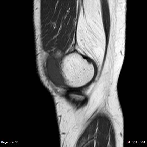
File:Anterior cruciate ligament tear (Radiopaedia 70783-80964 Sagittal T1 5).jpg
Jump to navigation
Jump to search
Anterior_cruciate_ligament_tear_(Radiopaedia_70783-80964_Sagittal_T1_5).jpg (560 × 560 pixels, file size: 24 KB, MIME type: image/jpeg)
Summary:
| Description |
|
| Date | Published: 16th Sep 2019 |
| Source | https://radiopaedia.org/cases/anterior-cruciate-ligament-tear-6 |
| Author | Ahmed Nafea |
| Permission (Permission-reusing-text) |
http://creativecommons.org/licenses/by-nc-sa/3.0/ |
Licensing:
Attribution-NonCommercial-ShareAlike 3.0 Unported (CC BY-NC-SA 3.0)
File history
Click on a date/time to view the file as it appeared at that time.
| Date/Time | Thumbnail | Dimensions | User | Comment | |
|---|---|---|---|---|---|
| current | 15:29, 8 May 2021 |  | 560 × 560 (24 KB) | Fæ (talk | contribs) | Radiopaedia project rID:70783 (batch #2202-71 C5) |
You cannot overwrite this file.
File usage
There are no pages that use this file.
