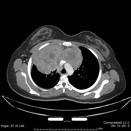
File:Anterior mediastinal B cell Lymphoma (Radiopaedia 50677-56114 A 24).jpg
Jump to navigation
Jump to search
Anterior_mediastinal_B_cell_Lymphoma_(Radiopaedia_50677-56114_A_24).jpg (512 × 512 pixels, file size: 28 KB, MIME type: image/jpeg)
Summary:
| Description |
|
| Date | Published: 30th Aug 2017 |
| Source | https://radiopaedia.org/cases/anterior-mediastinal-b-cell-lymphoma |
| Author | Abdallah Alqudah |
| Permission (Permission-reusing-text) |
http://creativecommons.org/licenses/by-nc-sa/3.0/ |
Licensing:
Attribution-NonCommercial-ShareAlike 3.0 Unported (CC BY-NC-SA 3.0)
File history
Click on a date/time to view the file as it appeared at that time.
| Date/Time | Thumbnail | Dimensions | User | Comment | |
|---|---|---|---|---|---|
| current | 07:12, 9 May 2021 |  | 512 × 512 (28 KB) | Fæ (talk | contribs) | Radiopaedia project rID:50677 (batch #2275-24 A24) |
You cannot overwrite this file.
File usage
The following page uses this file:
