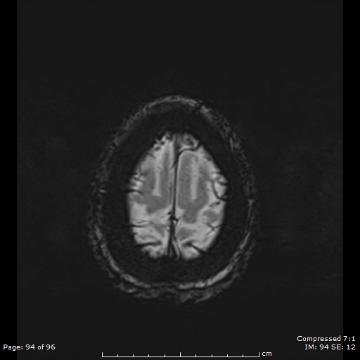
File:Anterior temporal lobe perivascular space (Radiopaedia 88283-104914 Axial SWI 63).jpg
Jump to navigation
Jump to search
Anterior_temporal_lobe_perivascular_space_(Radiopaedia_88283-104914_Axial_SWI_63).jpg (512 × 512 pixels, file size: 62 KB, MIME type: image/jpeg)
Summary:
| Description |
|
| Date | Published: 3rd Apr 2021 |
| Source | https://radiopaedia.org/cases/anterior-temporal-lobe-perivascular-space-4 |
| Author | Eid Kakish |
| Permission (Permission-reusing-text) |
http://creativecommons.org/licenses/by-nc-sa/3.0/ |
Licensing:
Attribution-NonCommercial-ShareAlike 3.0 Unported (CC BY-NC-SA 3.0)
File history
Click on a date/time to view the file as it appeared at that time.
| Date/Time | Thumbnail | Dimensions | User | Comment | |
|---|---|---|---|---|---|
| current | 21:03, 9 May 2021 |  | 512 × 512 (62 KB) | Fæ (talk | contribs) | Radiopaedia project rID:88283 (batch #2375-339 K63) |
You cannot overwrite this file.
File usage
The following page uses this file:
