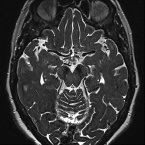
File:Anterior temporal pole cysts (Radiopaedia 46629-51102 Axial 51).png
Jump to navigation
Jump to search
Anterior_temporal_pole_cysts_(Radiopaedia_46629-51102_Axial_51).png (512 × 512 pixels, file size: 147 KB, MIME type: image/png)
Summary:
| Description |
|
| Date | Published: 14th Jul 2016 |
| Source | https://radiopaedia.org/cases/anterior-temporal-pole-cysts |
| Author | Frank Gaillard |
| Permission (Permission-reusing-text) |
http://creativecommons.org/licenses/by-nc-sa/3.0/ |
Licensing:
Attribution-NonCommercial-ShareAlike 3.0 Unported (CC BY-NC-SA 3.0)
File history
Click on a date/time to view the file as it appeared at that time.
| Date/Time | Thumbnail | Dimensions | User | Comment | |
|---|---|---|---|---|---|
| current | 22:44, 9 May 2021 |  | 512 × 512 (147 KB) | Fæ (talk | contribs) | Radiopaedia project rID:46629 (batch #2377-107 B51) |
You cannot overwrite this file.
File usage
The following page uses this file:
