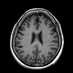
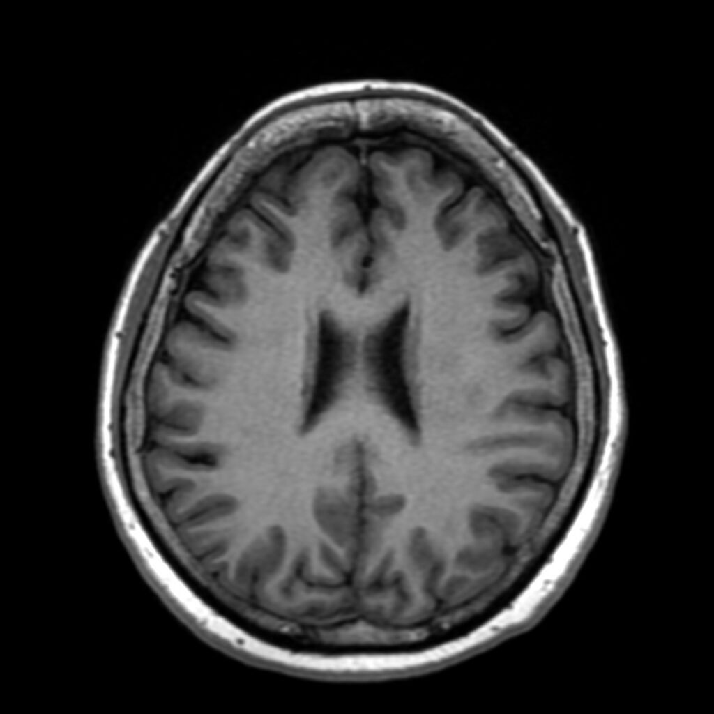
File:Anti-MOG encephalomyelitis (Radiopaedia 73831-84646 Axial T1 33).jpg
Jump to navigation
Jump to search

Size of this preview: 600 × 600 pixels. Other resolutions: 240 × 240 pixels | 480 × 480 pixels | 768 × 768 pixels | 1,024 × 1,024 pixels | 1,400 × 1,400 pixels.
Original file (1,400 × 1,400 pixels, file size: 162 KB, MIME type: image/jpeg)
Summary:
| Description |
|
| Date | Published: 24th Jan 2020 |
| Source | https://radiopaedia.org/cases/anti-mog-encephalomyelitis-1 |
| Author | Grace Carpenter |
| Permission (Permission-reusing-text) |
http://creativecommons.org/licenses/by-nc-sa/3.0/ |
Licensing:
Attribution-NonCommercial-ShareAlike 3.0 Unported (CC BY-NC-SA 3.0)
File history
Click on a date/time to view the file as it appeared at that time.
| Date/Time | Thumbnail | Dimensions | User | Comment | |
|---|---|---|---|---|---|
| current | 09:00, 10 May 2021 | | 1,400 × 1,400 (162 KB) | Fæ (talk | contribs) | Radiopaedia project rID:73831 (batch #2408-33 A33) |
You cannot overwrite this file.
File usage
The following page uses this file:
.jpg&oldid=334090)