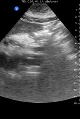
File:Aortic Dissection (Radiopaedia 85272-100839 C 1).mp4
Jump to navigation
Jump to search
Size of this JPG preview of this MP4 file: 404 × 599 pixels. Other resolutions: 162 × 240 pixels | 323 × 479 pixels | 728 × 1,080 pixels.
Original file (MP4 audio/video file, h.264, length 29 s, 728 × 1,080 pixels, 2.35 Mbps overall, file size: 8.08 MB)
Summary:
| Description |
|
| Date | 20 Dec 2020 |
| Source | Aortic dissection |
| Author | David Carroll |
| Permission (Permission-reusing-text) |
http://creativecommons.org/licenses/by-nc-sa/3.0/ |
Licensing:
Attribution-NonCommercial-ShareAlike 3.0 Unported (CC BY-NC-SA 3.0)
| This file is ineligible for copyright and therefore in the public domain, because it is a technical image created as part of a standard medical diagnostic procedure. No creative element rising above the threshold of originality was involved in its production.
|
 |
File history
Click on a date/time to view the file as it appeared at that time.
| Date/Time | Thumbnail | Dimensions | User | Comment | |
|---|---|---|---|---|---|
| current | 13:42, 11 May 2021 | 29 s, 728 × 1,080 (8.08 MB) | Fæ (talk | contribs) | Radiopaedia project rID:85272 (batch #2472-3 C1) |
You cannot overwrite this file.
File usage
There are no pages that use this file.
Transcode status
Update transcode status| Format | Bitrate | Download | Status | Encode time |
|---|---|---|---|---|
| VP9 1080P | 290 kbps | Download file | Completed 14:49, 11 May 2021 | 1 min 30 s |
| VP9 720P | 363 kbps | Download file | Completed 14:50, 11 May 2021 | 1 min 20 s |
| VP9 480P | 574 kbps | Download file | Completed 13:45, 11 May 2021 | 1 min 17 s |
| VP9 360P | 300 kbps | Download file | Completed 13:43, 11 May 2021 | 52 s |
| VP9 240P | 157 kbps | Download file | Completed 13:43, 11 May 2021 | 26 s |
| VP9 180P | 100 kbps | Download file | Completed 13:43, 11 May 2021 | 19 s |
| VP9 120P | 49 kbps | Download file | Completed 13:43, 11 May 2021 | 10 s |