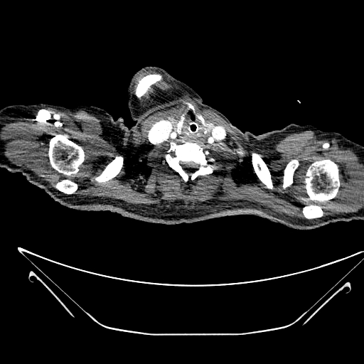
File:Aortic arch aneurysm (Radiopaedia 84109-99365 B 3).jpg

From NC Commons
Jump to navigation Jump to search

Aortic_arch_aneurysm_(Radiopaedia_84109-99365_B_3).jpg(512 × 512 pixels, file size: 92 KB, MIME type: image/jpeg)

Summary:

Description
  • Radiopaedia case ID: 84109
  • Image ID: 53869397
  • Image stack position: 3/399
  • Plane projection: Axial
  • Aux modality: C+ arterial phase
  • Study findings: Precontrast, a large well-defined rounded mainly superior mediastinal relatively homogenous non-calcified lesion is seen in direct contact and inseparable from the right wall of the aortic arch with an apparent continuity through a narrow-necked wall defect. Eccentric mural hyperdensity is seen along the right lateral wall of the lesion. No wall calcification is noted. Aortic arch mural calcification is noted. Post-contrast, there is contrast opacification within the described lesion similar to the adjacent aortic arch with a direct connection between them through a narrow-neck representing a large saccular aortic arch aneurysm. Mural thrombus is seen within the aneurysmal sac. Significant mass-effect is noted on the trachea and esophagus with deviation to the right side. Significant narrowing with incomplete encasement of the trachea and proximal main bronchi. Possible dissection flap of the most proximal left subclavian artery. No evidence of aneurysmal dilatation of ascending aorta, aortic arch branches, and descending aorta. Mild pericardial and bilateral pleural effusions with slightly hyperattenuating fluid representing hemopericardium and hemothorax likely from a leak. Multiple small consolidative opacities noted in the right lower lobe suggestive of an infectious process. Multiple enlarged subcarinal lymph nodes. Conclusion: Appearances are in keeping with a large aortic arch aneurysm with mural thrombus associated with hemopericardium and hemothorax concerning for a leak.
  • Modality: CT
  • System: Chest
  • Findings: The mediastinum is widened with a large mainly superior mediastinal non-calcified rounded opacity deviating the trachea to the right side.
Date Published: 17th Feb 2021
Source https://radiopaedia.org/cases/aortic-arch-aneurysm-4
Author Fadi Aidi
Permission
(Permission-reusing-text)
http://creativecommons.org/licenses/by-nc-sa/3.0/

Licensing:

Attribution-NonCommercial-ShareAlike 3.0 Unported (CC BY-NC-SA 3.0)

File history

Click on a date/time to view the file as it appeared at that time.

Date/TimeThumbnailDimensionsUserComment
current19:20, 10 May 2021Thumbnail for version as of 19:20, 10 May 2021512 × 512 (92 KB) (talk | contribs)Radiopaedia project rID:84109 (batch #2439-402 B3)

The following page uses this file: