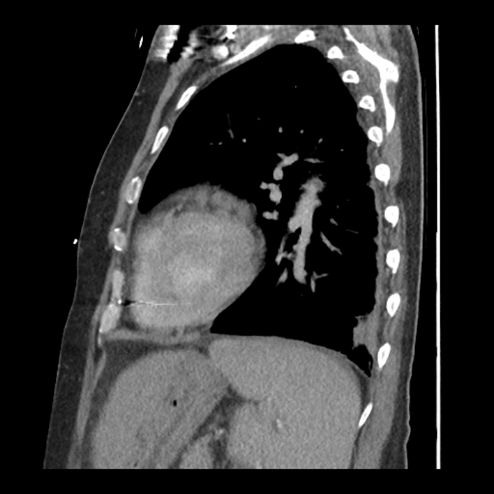
File:Aortic arch graft infection (FDG PET-CT) (Radiopaedia 71975-82437 C 50).jpg
Jump to navigation
Jump to search
Size of this preview: 600 × 600 pixels. Other resolutions: 240 × 240 pixels | 480 × 480 pixels | 708 × 708 pixels.
Original file (708 × 708 pixels, file size: 139 KB, MIME type: image/jpeg)
Summary:
| Description |
|
| Date | Published: 1st Nov 2019 |
| Source | https://radiopaedia.org/cases/aortic-arch-graft-infection-fdg-pet-ct |
| Author | A A |
| Permission (Permission-reusing-text) |
http://creativecommons.org/licenses/by-nc-sa/3.0/ |
Licensing:
Attribution-NonCommercial-ShareAlike 3.0 Unported (CC BY-NC-SA 3.0)
File history
Click on a date/time to view the file as it appeared at that time.
| Date/Time | Thumbnail | Dimensions | User | Comment | |
|---|---|---|---|---|---|
| current | 23:09, 10 May 2021 | | 708 × 708 (139 KB) | Fæ (talk | contribs) | Radiopaedia project rID:71975 (batch #2441-185 C50) |
You cannot overwrite this file.
File usage
The following page uses this file:
_(Radiopaedia_71975-82437_C_50).jpg&oldid=339956)