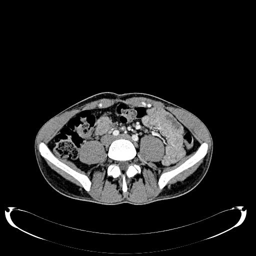
File:Aortic coarctation (Radiopaedia 53947-60320 A 253).jpg
Jump to navigation
Jump to search
Aortic_coarctation_(Radiopaedia_53947-60320_A_253).jpg (512 × 512 pixels, file size: 24 KB, MIME type: image/jpeg)
Summary:
| Description |
|
| Date | Published: 27th Jun 2017 |
| Source | https://radiopaedia.org/cases/aortic-coarctation-2 |
| Author | Arkadi Tadevosyan |
| Permission (Permission-reusing-text) |
http://creativecommons.org/licenses/by-nc-sa/3.0/ |
Licensing:
Attribution-NonCommercial-ShareAlike 3.0 Unported (CC BY-NC-SA 3.0)
File history
Click on a date/time to view the file as it appeared at that time.
| Date/Time | Thumbnail | Dimensions | User | Comment | |
|---|---|---|---|---|---|
| current | 04:11, 11 May 2021 |  | 512 × 512 (24 KB) | Fæ (talk | contribs) | Radiopaedia project rID:53947 (batch #2454-253 A253) |
You cannot overwrite this file.
File usage
The following page uses this file:
