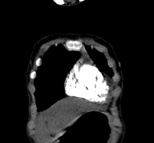
File:Aortic coarctation (Radiopaedia 69135-78920 C 6).jpg
Jump to navigation
Jump to search
Aortic_coarctation_(Radiopaedia_69135-78920_C_6).jpg (512 × 475 pixels, file size: 46 KB, MIME type: image/jpeg)
Summary:
| Description |
|
| Date | Published: 30th Jun 2019 |
| Source | https://radiopaedia.org/cases/aortic-coarctation-4 |
| Author | Fazel Rahman Faizi |
| Permission (Permission-reusing-text) |
http://creativecommons.org/licenses/by-nc-sa/3.0/ |
Licensing:
Attribution-NonCommercial-ShareAlike 3.0 Unported (CC BY-NC-SA 3.0)
File history
Click on a date/time to view the file as it appeared at that time.
| Date/Time | Thumbnail | Dimensions | User | Comment | |
|---|---|---|---|---|---|
| current | 02:21, 11 May 2021 |  | 512 × 475 (46 KB) | Fæ (talk | contribs) | Radiopaedia project rID:69135 (batch #2451-203 C6) |
You cannot overwrite this file.
File usage
The following page uses this file:
