File:Aortic dissection (Radiopaedia 25350-25604 E 10).jpg

Original file (1,024 × 1,024 pixels, file size: 127 KB, MIME type: image/jpeg)
Summary:
| Description |
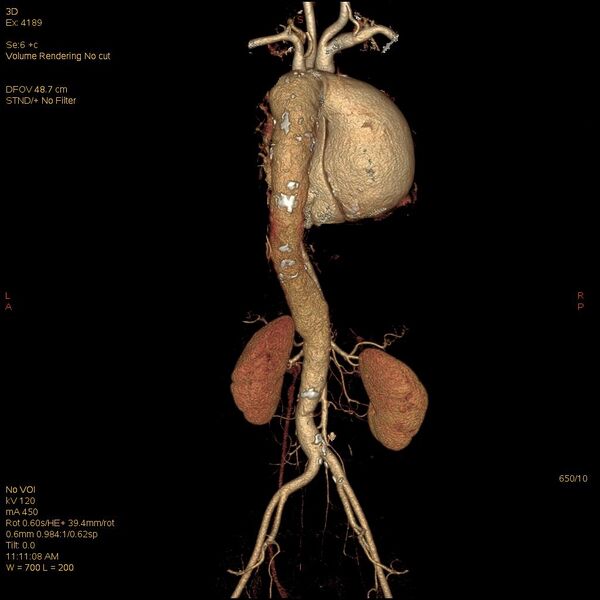
aorta, aortic arch and descending aorta. An intimal flap is seen extending from the aortic root proximally to below the origin of the renal arteries distally dividing the aortic lumen into true and false lumens (Stanford Type A and type I DeBakey classification). Right brachiocephalic, left common carotid, left subclavian, superior mesenteric, inferior mesenteric and bilateral renal arteries are seen arising from true lumen. There is significant narrowing at the origin of celiac trunk of abdominal aorta. Atherosclerotic wall calcification is noted at origin of left renal artery. Cardiomegaly with dilatation of all cardiac chambers is noted. |
| Date | Published: 21st Oct 2013 |
| Source | https://radiopaedia.org/cases/aortic-dissection-13 |
| Author | Prashant Mudgal |
| Permission (Permission-reusing-text) |
http://creativecommons.org/licenses/by-nc-sa/3.0/ |
Licensing:
Attribution-NonCommercial-ShareAlike 3.0 Unported (CC BY-NC-SA 3.0)
File history
Click on a date/time to view the file as it appeared at that time.
| Date/Time | Thumbnail | Dimensions | User | Comment | |
|---|---|---|---|---|---|
| current | 12:49, 11 May 2021 | | 1,024 × 1,024 (127 KB) | Fæ (talk | contribs) | Radiopaedia project rID:25350 (batch #2469-173 E10) |
You cannot overwrite this file.
File usage
There are no pages that use this file.
.jpg&oldid=345168)