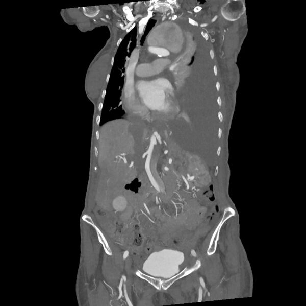
File:Aortic dissection (Radiopaedia 68763-78691 B 14).jpeg

From NC Commons
Jump to navigation Jump to search

Original file(845 × 844 pixels, file size: 99 KB, MIME type: image/jpeg)

Summary:

Description
  • Radiopaedia case ID: 68763
  • Image ID: 49686101
  • Image stack position: 14/34
  • Plane projection: Coronal
  • Aux modality: C+ arterial phase
  • Study findings: There is a Type B aortic dissection extending from the distal aortic arch into the left common iliac artery associated with a large aneurysm of the proximal descending thoracic aorta. There is no active contrast leak identified. As already noted in the prior chest radiograph, there is a large left-sided pleural effusion with nearly complete collapse and consolidation of the left lung. This is highly likely to be secondary to rupture/leaking of the aneurysm. The major mesenteric vessels are noted to originate from the true lumen. The left renal artery shows reduced contrast filling and the left kidney is ischemic.
  • Modality: CT
  • System: Cardiac
  • Findings: There is opacification of the lower two-thirds of the left hemithorax indicative of large pleural effusion. This is associated with a positive mass effect with displacement of the mediastinum to the right. There is an impression of a large superior mediastinal mass on the left with tracheal deviation to the right. There is evidence of a small right-sided pleural effusion.
Date Published: 21st Jun 2019
Source https://radiopaedia.org/cases/aortic-dissection-34
Author Devanshi Pathania
Permission
(Permission-reusing-text)
http://creativecommons.org/licenses/by-nc-sa/3.0/

Licensing:

Attribution-NonCommercial-ShareAlike 3.0 Unported (CC BY-NC-SA 3.0)

File history

Click on a date/time to view the file as it appeared at that time.

Date/TimeThumbnailDimensionsUserComment
current11:36, 11 May 2021Thumbnail for version as of 11:36, 11 May 2021845 × 844 (99 KB) (talk | contribs)Radiopaedia project rID:68763 (batch #2464-69 B14)

There are no pages that use this file.

Metadata