File:Aortic dissection - Stanford A - DeBakey I (Radiopaedia 23469-23551 Axial MRA 11).jpg
Aortic_dissection_-_Stanford_A_-_DeBakey_I_(Radiopaedia_23469-23551_Axial_MRA_11).jpg (512 × 512 pixels, file size: 63 KB, MIME type: image/jpeg)
Summary:
| Description |
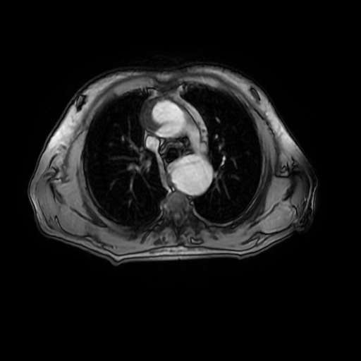
arising in the ascending aorta shortly distal to the aortic root and spirals along the whole aorta forming large false lumen mainly posterior to and compressing the true lumen which appears ovoid in cross-section. The flap extends into the upper scanned common iliac arteries. It shows fenestration at the proximal aortic arch and at just proximal to the aortic bifurcation. The aorta overall is seen dilated with largest diameter is at the distal thoracic descending aorta measuring 5.1 cm. The false lumen shows non-uniform partial thrombosis, thickest at the distal descending thoracic aorta reducing its diameter by 50%. The narrowest point of the true lumen is at level of diaphragmatic hiatus and measures 2.9 X 1 cm. The celiac trunk mesenteric artery and both renal arteries are seen arising from the true lumen; with the renal arteries are seen relatively splayed by the displaced dilated aorta. |
| Date | Published: 18th Jun 2013 |
| Source | https://radiopaedia.org/cases/aortic-dissection-stanford-a-debakey-i-1 |
| Author | Mohammad A. ElBeialy |
| Permission (Permission-reusing-text) |
http://creativecommons.org/licenses/by-nc-sa/3.0/ |
Licensing:
Attribution-NonCommercial-ShareAlike 3.0 Unported (CC BY-NC-SA 3.0)
File history
Click on a date/time to view the file as it appeared at that time.
| Date/Time | Thumbnail | Dimensions | User | Comment | |
|---|---|---|---|---|---|
| current | 20:31, 11 May 2021 | | 512 × 512 (63 KB) | Fæ (talk | contribs) | Radiopaedia project rID:23469 (batch #2488-71 C11) |
You cannot overwrite this file.
.jpg&oldid=1674409)