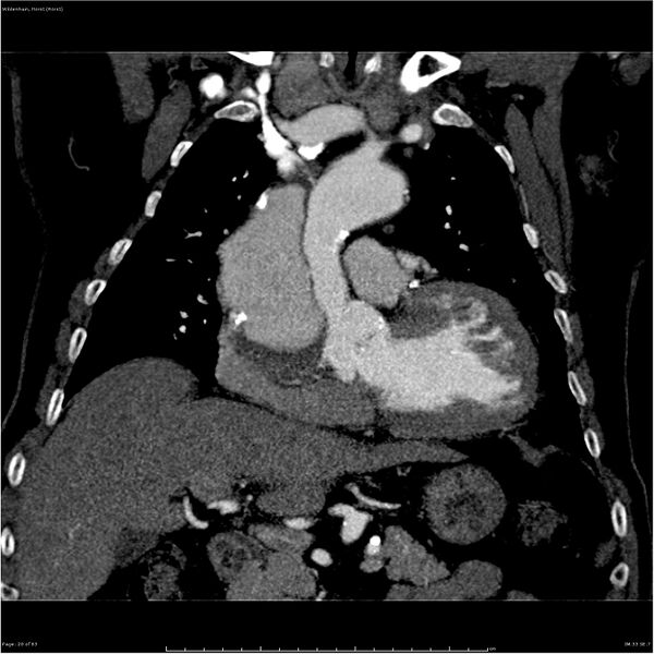
File:Aortic dissection - Stanford type A (Radiopaedia 26183-26315 A 20).jpg
Jump to navigation
Jump to search

Size of this preview: 600 × 600 pixels. Other resolutions: 240 × 240 pixels | 480 × 480 pixels | 768 × 768 pixels | 1,024 × 1,024 pixels | 1,733 × 1,733 pixels.
Original file (1,733 × 1,733 pixels, file size: 771 KB, MIME type: image/jpeg)
Summary:
| Description |
|
| Date | Published: 4th Dec 2013 |
| Source | https://radiopaedia.org/cases/aortic-dissection-stanford-type-a-13 |
| Author | Henry Knipe |
| Permission (Permission-reusing-text) |
http://creativecommons.org/licenses/by-nc-sa/3.0/ |
Licensing:
Attribution-NonCommercial-ShareAlike 3.0 Unported (CC BY-NC-SA 3.0)
File history
Click on a date/time to view the file as it appeared at that time.
| Date/Time | Thumbnail | Dimensions | User | Comment | |
|---|---|---|---|---|---|
| current | 23:40, 11 May 2021 |  | 1,733 × 1,733 (771 KB) | Fæ (talk | contribs) | Radiopaedia project rID:26183 (batch #2495-20 A20) |
You cannot overwrite this file.
File usage
The following page uses this file: