File:Aortic dissection - Stanford type B (Radiopaedia 50171-55512 A 59).png
Jump to navigation
Jump to search
No higher resolution available.
Aortic_dissection_-_Stanford_type_B_(Radiopaedia_50171-55512_A_59).png (512 × 512 pixels, file size: 86 KB, MIME type: image/png)
Summary:
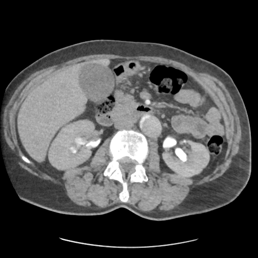
| Description |
|
| Date | Published: 22nd Dec 2016 |
| Source | https://radiopaedia.org/cases/aortic-dissection-stanford-type-b-5 |
| Author | Henry Knipe |
| Permission (Permission-reusing-text) |
http://creativecommons.org/licenses/by-nc-sa/3.0/ |
Licensing:
Attribution-NonCommercial-ShareAlike 3.0 Unported (CC BY-NC-SA 3.0)
File history
Click on a date/time to view the file as it appeared at that time.
| Date/Time | Thumbnail | Dimensions | User | Comment | |
|---|---|---|---|---|---|
| current | 03:32, 12 May 2021 | | 512 × 512 (86 KB) | Fæ (talk | contribs) | Radiopaedia project rID:50171 (batch #2502-59 A59) |
You cannot overwrite this file.
File usage
The following page uses this file:
.png&oldid=350395)