File:Aortic intramural hematoma (Radiopaedia 34260-35540 B 82).png

From NC Commons
Jump to navigation Jump to search

Aortic_intramural_hematoma_(Radiopaedia_34260-35540_B_82).png(512 × 512 pixels, file size: 152 KB, MIME type: image/png)

Summary:

Description
  • Radiopaedia case ID: 34260
  • Image ID: 11020381
  • Image stack position: 82/96
  • Plane projection: Axial
  • Aux modality: C+ arterial phase
  • Modality: CT
  • System: Vascular
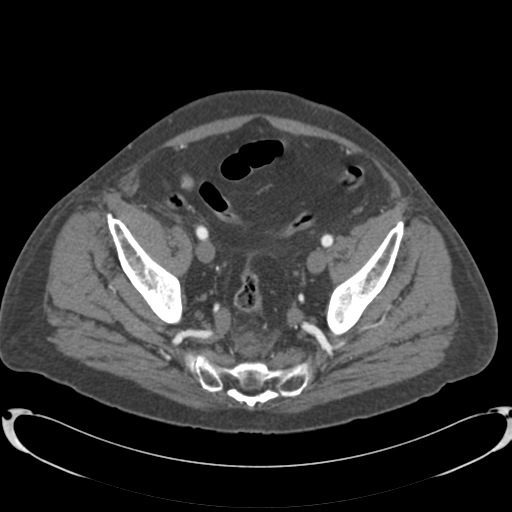
  • Findings: There is mural thickening of the aorta extending from the arch (at the origin of the subclavian artery) to just above the renal artery. This is predominantly along the superior wall of the arch and along the left posterolateral aspect in the thoracic descending aorta. The mural thickening is high density on the precontrast images. There are pockets of contrast at various points within the mural thickening. A discrete intimal flap is not demonstrated except at the inferior extent of the mural thickening where there is the suggestion of a short segment flap. There was a tiny focus of mural enhancement on the previous study at this site. Maximum thoracic aortic diameter 3.8cm. There is also an 4.6cm infrarenal fusiform abdominal aortic aneurysm extending to the bifurcation of the aorta with a 2.6cm neck. Scattered calcific atheroma. Conclusion: 1. The findings are consistent with aortic intramural hematoma (Type B).
Date Published: 12th Feb 2015
Source https://radiopaedia.org/cases/aortic-intramural-haematoma-3
Author RMH Core Conditions
Permission
(Permission-reusing-text)
http://creativecommons.org/licenses/by-nc-sa/3.0/

Licensing:

Attribution-NonCommercial-ShareAlike 3.0 Unported (CC BY-NC-SA 3.0)

File history

Click on a date/time to view the file as it appeared at that time.

Date/TimeThumbnailDimensionsUserComment
current01:49, 18 May 2021Thumbnail for version as of 01:49, 18 May 2021512 × 512 (152 KB) (talk | contribs)Radiopaedia project rID:34260 (batch #2518-139 B82)

The following page uses this file:

Metadata