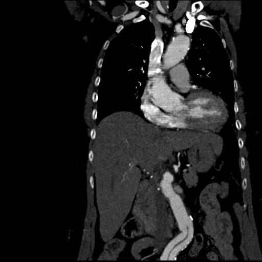
File:Aortic intramural hematoma from penetrating atherosclerotic ulcer (Radiopaedia 31137-31836 C 26).jpg
Jump to navigation
Jump to search
No higher resolution available.
Aortic_intramural_hematoma_from_penetrating_atherosclerotic_ulcer_(Radiopaedia_31137-31836_C_26).jpg (512 × 512 pixels, file size: 27 KB, MIME type: image/jpeg)
Summary:
| Description |
|
| Date | Published: 22nd Sep 2014 |
| Source | https://radiopaedia.org/cases/aortic-intramural-haematoma-from-penetrating-atherosclerotic-ulcer |
| Author | RMH Core Conditions |
| Permission (Permission-reusing-text) |
http://creativecommons.org/licenses/by-nc-sa/3.0/ |
Licensing:
Attribution-NonCommercial-ShareAlike 3.0 Unported (CC BY-NC-SA 3.0)
File history
Click on a date/time to view the file as it appeared at that time.
| Date/Time | Thumbnail | Dimensions | User | Comment | |
|---|---|---|---|---|---|
| current | 05:16, 18 May 2021 | | 512 × 512 (27 KB) | Fæ (talk | contribs) | Radiopaedia project rID:31137 (batch #2524-184 C26) |
You cannot overwrite this file.
File usage
The following page uses this file:
.jpg&oldid=354616)