File:Aortic intramural hematoma with dissection and intramural blood pool (Radiopaedia 77373-89491 D 53).jpg
Jump to navigation
Jump to search
No higher resolution available.
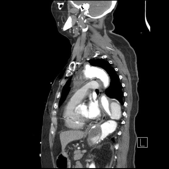
Aortic_intramural_hematoma_with_dissection_and_intramural_blood_pool_(Radiopaedia_77373-89491_D_53).jpg (548 × 548 pixels, file size: 106 KB, MIME type: image/jpeg)
Summary:
| Description |
|
| Date | Published: 4th Jun 2020 |
| Source | https://radiopaedia.org/cases/aortic-intramural-haematoma-with-dissection-and-intramural-blood-pool |
| Author | Craig Hacking |
| Permission (Permission-reusing-text) |
http://creativecommons.org/licenses/by-nc-sa/3.0/ |
Licensing:
Attribution-NonCommercial-ShareAlike 3.0 Unported (CC BY-NC-SA 3.0)
File history
Click on a date/time to view the file as it appeared at that time.
| Date/Time | Thumbnail | Dimensions | User | Comment | |
|---|---|---|---|---|---|
| current | 07:56, 18 May 2021 | | 548 × 548 (106 KB) | Fæ (talk | contribs) | Radiopaedia project rID:77373 (batch #2526-352 D53) |
You cannot overwrite this file.
File usage
The following page uses this file:
.jpg&oldid=355686)