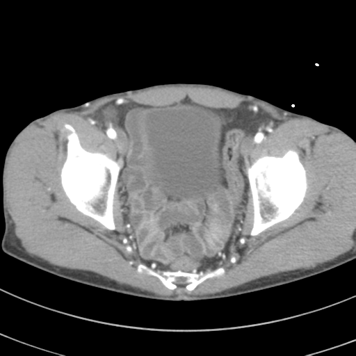
File:Aortoiliac occlusive disease (Radiopaedia 57950-64931 A 64).png
Jump to navigation
Jump to search
Aortoiliac_occlusive_disease_(Radiopaedia_57950-64931_A_64).png (512 × 512 pixels, file size: 69 KB, MIME type: image/png)
Summary:
| Description |
|
| Date | Published: 3rd Mar 2018 |
| Source | https://radiopaedia.org/cases/aortoiliac-occlusive-disease-7 |
| Author | Henry Knipe |
| Permission (Permission-reusing-text) |
http://creativecommons.org/licenses/by-nc-sa/3.0/ |
Licensing:
Attribution-NonCommercial-ShareAlike 3.0 Unported (CC BY-NC-SA 3.0)
File history
Click on a date/time to view the file as it appeared at that time.
| Date/Time | Thumbnail | Dimensions | User | Comment | |
|---|---|---|---|---|---|
| current | 02:02, 19 May 2021 |  | 512 × 512 (69 KB) | Fæ (talk | contribs) | Radiopaedia project rID:57950 (batch #2567-64 A64) |
You cannot overwrite this file.
File usage
The following page uses this file:
