File:Apert syndrome (Radiopaedia 6357-7723 A 1).jpg

From NC Commons
Jump to navigation Jump to search

Apert_syndrome_(Radiopaedia_6357-7723_A_1).jpg(800 × 600 pixels, file size: 93 KB, MIME type: image/jpeg)

Summary:

Description
  • Radiopaedia case ID: 6357
  • Image ID: 25266
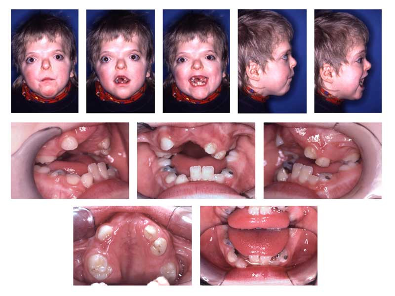
  • Study findings: Figure 1 Age 8.8. Extraorally: Hypertelorism, vertical excess of the lower third of the face, trapezoidal upper lip, forced lip closure possible, but difficult. Intraorally: Dentitio tarda, crowding, Angle Class III with dental compensation by retrusion of lower incisors, circular open bite with unilateral crossbite. Reference: Hohoff Ariane, Ulrich Joos, Ulrich Meyer et al. “The spectrum of Apert syndrome: phenotype, particularities in orthodontic treatment, and characteristics of orthognathic surgery.” Head & Face Medicine 3, no. 1 (2007): 10.
  • Modality: Photo
  • System: Musculoskeletal
  • Findings: Figure 2: OPG Age 13.3. After 22 months in braces and genioplasty. Left: Panoramic radiograph does not reveal root resorption. Teeth 27, 38, and 48 were removed at time of genioplasty. Right: Superimposition of lateral ceph before treatment with the fixed appliance (blue) (age 11.4) and after genioplasty.
Date Published: 11th Jun 2009
Source https://radiopaedia.org/cases/apert-syndrome-2
Author Frank Gaillard
Permission
(Permission-reusing-text)
http://creativecommons.org/licenses/by-nc-sa/3.0/

Licensing:

Attribution-NonCommercial-ShareAlike 3.0 Unported (CC BY-NC-SA 3.0)

File history

Click on a date/time to view the file as it appeared at that time.

Date/TimeThumbnailDimensionsUserComment
current07:04, 19 May 2021Thumbnail for version as of 07:04, 19 May 2021800 × 600 (93 KB) (talk | contribs)Radiopaedia project rID:6357 (batch #2584-1 A1)

There are no pages that use this file.