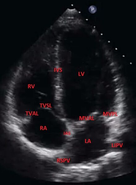
File:Apical 4 chamber view - normal (transthoracic echocardiography) (Radiopaedia 62756).jpg
Jump to navigation
Jump to search

Size of this preview: 440 × 599 pixels. Other resolutions: 176 × 240 pixels | 482 × 656 pixels.
Original file (482 × 656 pixels, file size: 47 KB, MIME type: image/jpeg)
Summary:
| Description |
|
| Date | 28 Aug 2018 |
| Source | Apical 4 chamber view - normal (transthoracic echocardiography) |
| Author | David Carroll |
| Permission (Permission-reusing-text) |
http://creativecommons.org/licenses/by-nc-sa/3.0/ |
Licensing:
Attribution-NonCommercial-ShareAlike 3.0 Unported (CC BY-NC-SA 3.0)
| This file is ineligible for copyright and therefore in the public domain, because it is a technical image created as part of a standard medical diagnostic procedure. No creative element rising above the threshold of originality was involved in its production.
|
 |
File history
Click on a date/time to view the file as it appeared at that time.
| Date/Time | Thumbnail | Dimensions | User | Comment | |
|---|---|---|---|---|---|
| current | 22:56, 18 March 2021 | | 482 × 656 (47 KB) | Fæ (talk | contribs) | Radiopaedia project rID:62756 (batch #2540) |
You cannot overwrite this file.
File usage
The following page uses this file:
_(Radiopaedia_62756).jpg&oldid=9291491)