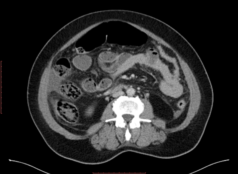
File:Appedicular abscess (Radiopaedia 79555-92727 B 30).jpg
Jump to navigation
Jump to search
Appedicular_abscess_(Radiopaedia_79555-92727_B_30).jpg (792 × 582 pixels, file size: 160 KB, MIME type: image/jpeg)
Summary:
| Description |
|
| Date | Published: 1st Jul 2020 |
| Source | https://radiopaedia.org/cases/appedicular-abscess |
| Author | Dr Husam Hussein Yaseen |
| Permission (Permission-reusing-text) |
http://creativecommons.org/licenses/by-nc-sa/3.0/ |
Licensing:
Attribution-NonCommercial-ShareAlike 3.0 Unported (CC BY-NC-SA 3.0)
File history
Click on a date/time to view the file as it appeared at that time.
| Date/Time | Thumbnail | Dimensions | User | Comment | |
|---|---|---|---|---|---|
| current | 14:04, 19 May 2021 |  | 792 × 582 (160 KB) | Fæ (talk | contribs) | Radiopaedia project rID:79555 (batch #2632-103 B30) |
You cannot overwrite this file.
File usage
The following page uses this file:
