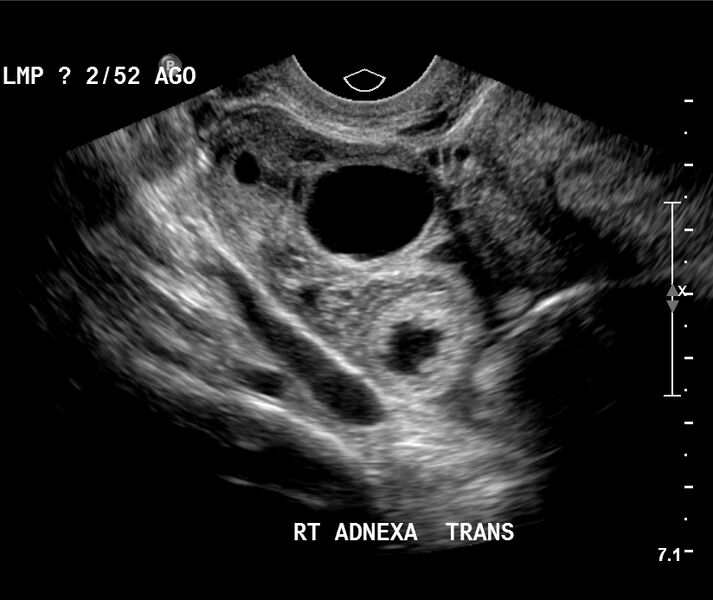
File:Appendiceal abscess (Radiopaedia 13096-13153 A 3).jpg
Jump to navigation
Jump to search

Size of this preview: 713 × 600 pixels. Other resolutions: 285 × 240 pixels | 571 × 480 pixels | 824 × 693 pixels.
Original file (824 × 693 pixels, file size: 194 KB, MIME type: image/jpeg)
Summary:
| Description |
|
| Date | Published: 1st Mar 2011 |
| Source | https://radiopaedia.org/cases/appendiceal-abscess |
| Author | Alexandra Stanislavsky |
| Permission (Permission-reusing-text) |
http://creativecommons.org/licenses/by-nc-sa/3.0/ |
Licensing:
Attribution-NonCommercial-ShareAlike 3.0 Unported (CC BY-NC-SA 3.0)
File history
Click on a date/time to view the file as it appeared at that time.
| Date/Time | Thumbnail | Dimensions | User | Comment | |
|---|---|---|---|---|---|
| current | 14:16, 19 May 2021 |  | 824 × 693 (194 KB) | Fæ (talk | contribs) | Radiopaedia project rID:13096 (batch #2633-3 A3) |
You cannot overwrite this file.
File usage
There are no pages that use this file.