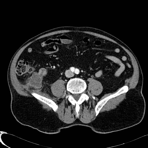
File:Appendiceal adenocarcinoma complicated by retroperitoneal abscess (Radiopaedia 58007-65040 Axial 1).jpg
Jump to navigation
Jump to search
No higher resolution available.
Appendiceal_adenocarcinoma_complicated_by_retroperitoneal_abscess_(Radiopaedia_58007-65040_Axial_1).jpg (512 × 512 pixels, file size: 62 KB, MIME type: image/jpeg)
Summary:
| Description |
|
| Date | Published: 26th Jan 2018 |
| Source | https://radiopaedia.org/cases/appendiceal-adenocarcinoma-complicated-by-retroperitoneal-abscess |
| Author | Francis Fortin |
| Permission (Permission-reusing-text) |
http://creativecommons.org/licenses/by-nc-sa/3.0/ |
Licensing:
Attribution-NonCommercial-ShareAlike 3.0 Unported (CC BY-NC-SA 3.0)
File history
Click on a date/time to view the file as it appeared at that time.
| Date/Time | Thumbnail | Dimensions | User | Comment | |
|---|---|---|---|---|---|
| current | 15:17, 19 May 2021 | | 512 × 512 (62 KB) | Fæ (talk | contribs) | Radiopaedia project rID:58007 (batch #2635-3 C1) |
You cannot overwrite this file.
File usage
The following file is a duplicate of this file (more details):
.jpg)
.jpg)
There are no pages that use this file.
.jpg&oldid=367755)