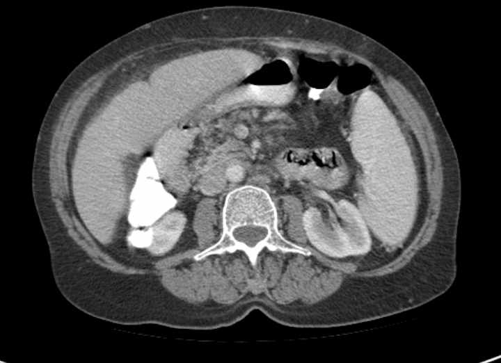
File:Appendiceal mucocele (Radiopaedia 73821-84638 A 47).jpg
Jump to navigation
Jump to search
No higher resolution available.
Appendiceal_mucocele_(Radiopaedia_73821-84638_A_47).jpg (720 × 522 pixels, file size: 32 KB, MIME type: image/jpeg)
Summary:
| Description |
|
| Date | Published: 23rd Jan 2020 |
| Source | https://radiopaedia.org/cases/appendiceal-mucocele-17 |
| Author | Yair Glick |
| Permission (Permission-reusing-text) |
http://creativecommons.org/licenses/by-nc-sa/3.0/ |
Licensing:
Attribution-NonCommercial-ShareAlike 3.0 Unported (CC BY-NC-SA 3.0)
File history
Click on a date/time to view the file as it appeared at that time.
| Date/Time | Thumbnail | Dimensions | User | Comment | |
|---|---|---|---|---|---|
| current | 23:39, 19 May 2021 | | 720 × 522 (32 KB) | Fæ (talk | contribs) | Radiopaedia project rID:73821 (batch #2648-47 A47) |
You cannot overwrite this file.
File usage
The following page uses this file:
.jpg&oldid=371048)