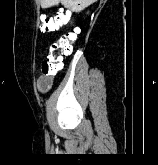
File:Appendiceal mucocele (Radiopaedia 83147-97518 D 19).jpg
Jump to navigation
Jump to search
Appendiceal_mucocele_(Radiopaedia_83147-97518_D_19).jpg (508 × 532 pixels, file size: 33 KB, MIME type: image/jpeg)
Summary:
| Description |
|
| Date | Published: 17th Oct 2020 |
| Source | https://radiopaedia.org/cases/appendiceal-mucocele-24 |
| Author | Mohammad Taghi Niknejad |
| Permission (Permission-reusing-text) |
http://creativecommons.org/licenses/by-nc-sa/3.0/ |
Licensing:
Attribution-NonCommercial-ShareAlike 3.0 Unported (CC BY-NC-SA 3.0)
File history
Click on a date/time to view the file as it appeared at that time.
| Date/Time | Thumbnail | Dimensions | User | Comment | |
|---|---|---|---|---|---|
| current | 23:19, 19 May 2021 |  | 508 × 532 (33 KB) | Fæ (talk | contribs) | Radiopaedia project rID:83147 (batch #2646-313 D19) |
You cannot overwrite this file.
File usage
The following page uses this file:
