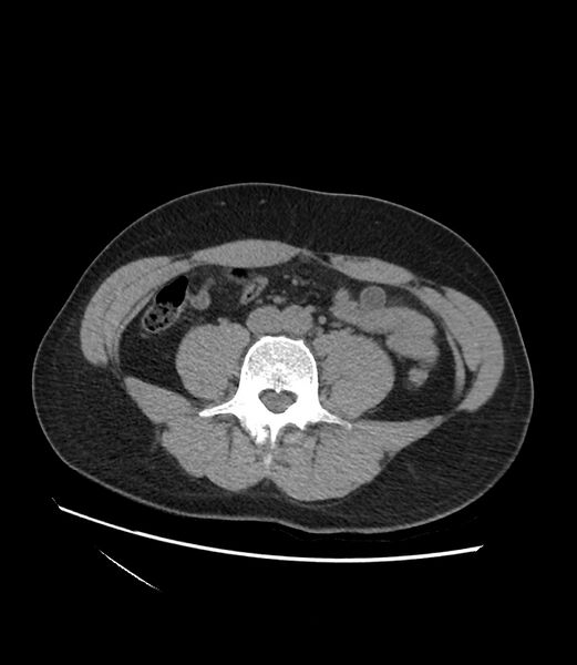
File:Appendiceal mucocele (Radiopaedia 86110-102060 Axial non-contrast 44).jpg
Jump to navigation
Jump to search

Size of this preview: 521 × 600 pixels. Other resolutions: 208 × 240 pixels | 417 × 480 pixels | 667 × 768 pixels | 890 × 1,024 pixels | 1,618 × 1,862 pixels.
Original file (1,618 × 1,862 pixels, file size: 209 KB, MIME type: image/jpeg)
Summary:
| Description |
|
| Date | Published: 20th Jan 2021 |
| Source | https://radiopaedia.org/cases/appendiceal-mucocele-26 |
| Author | Saad Ahmed Saad Hassan |
| Permission (Permission-reusing-text) |
http://creativecommons.org/licenses/by-nc-sa/3.0/ |
Licensing:
Attribution-NonCommercial-ShareAlike 3.0 Unported (CC BY-NC-SA 3.0)
File history
Click on a date/time to view the file as it appeared at that time.
| Date/Time | Thumbnail | Dimensions | User | Comment | |
|---|---|---|---|---|---|
| current | 02:27, 20 May 2021 |  | 1,618 × 1,862 (209 KB) | Fæ (talk | contribs) | Radiopaedia project rID:86110 (batch #2652-89 B44) |
You cannot overwrite this file.
File usage
The following page uses this file: