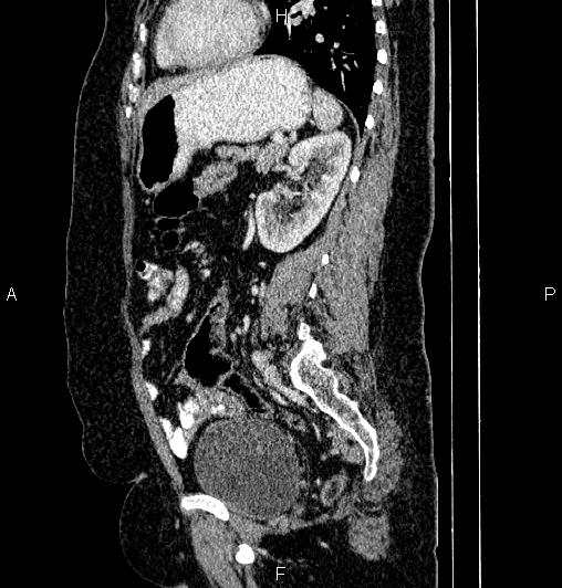
File:Appendiceal mucocele (Radiopaedia 86415-102460 E 62).jpg
Jump to navigation
Jump to search
Appendiceal_mucocele_(Radiopaedia_86415-102460_E_62).jpg (508 × 532 pixels, file size: 53 KB, MIME type: image/jpeg)
Summary:
| Description |
|
| Date | Published: 31st Jan 2021 |
| Source | https://radiopaedia.org/cases/appendiceal-mucocele-27 |
| Author | Mohammad Taghi Niknejad |
| Permission (Permission-reusing-text) |
http://creativecommons.org/licenses/by-nc-sa/3.0/ |
Licensing:
Attribution-NonCommercial-ShareAlike 3.0 Unported (CC BY-NC-SA 3.0)
File history
Click on a date/time to view the file as it appeared at that time.
| Date/Time | Thumbnail | Dimensions | User | Comment | |
|---|---|---|---|---|---|
| current | 03:53, 20 May 2021 |  | 508 × 532 (53 KB) | Fæ (talk | contribs) | Radiopaedia project rID:86415 (batch #2655-325 E62) |
You cannot overwrite this file.
File usage
The following page uses this file:
