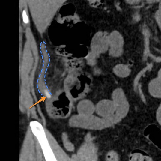
File:Appendicitis (Radiopaedia 12510-17135 A 1).jpg
Jump to navigation
Jump to search
Appendicitis_(Radiopaedia_12510-17135_A_1).jpg (537 × 537 pixels, file size: 48 KB, MIME type: image/jpeg)
Summary:
| Description |
|
| Date | Published: 7th Dec 2010 |
| Source | https://radiopaedia.org/cases/appendicitis-3 |
| Author | Frank Gaillard |
| Permission (Permission-reusing-text) |
http://creativecommons.org/licenses/by-nc-sa/3.0/ |
Licensing:
Attribution-NonCommercial-ShareAlike 3.0 Unported (CC BY-NC-SA 3.0)
File history
Click on a date/time to view the file as it appeared at that time.
| Date/Time | Thumbnail | Dimensions | User | Comment | |
|---|---|---|---|---|---|
| current | 10:40, 20 May 2021 |  | 537 × 537 (48 KB) | Fæ (talk | contribs) | Radiopaedia project rID:12510 (batch #2679-1 A1) |
You cannot overwrite this file.
File usage
There are no pages that use this file.
