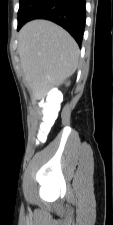
File:Appendicitis and incidental foregut duplication cyst (Radiopaedia 52962-58916 C 16).jpg
Jump to navigation
Jump to search
Size of this preview: 303 × 598 pixels. Other resolutions: 121 × 240 pixels | 389 × 768 pixels.
Original file (389 × 768 pixels, file size: 33 KB, MIME type: image/jpeg)
Summary:
| Description |
|
| Date | Published: 27th Apr 2017 |
| Source | https://radiopaedia.org/cases/appendicitis-and-incidental-foregut-duplication-cyst |
| Author | Yair Glick |
| Permission (Permission-reusing-text) |
http://creativecommons.org/licenses/by-nc-sa/3.0/ |
Licensing:
Attribution-NonCommercial-ShareAlike 3.0 Unported (CC BY-NC-SA 3.0)
File history
Click on a date/time to view the file as it appeared at that time.
| Date/Time | Thumbnail | Dimensions | User | Comment | |
|---|---|---|---|---|---|
| current | 14:04, 20 May 2021 | | 389 × 768 (33 KB) | Fæ (talk | contribs) | Radiopaedia project rID:52962 (batch #2690-184 C16) |
You cannot overwrite this file.
File usage
The following page uses this file:
.jpg&oldid=376438)