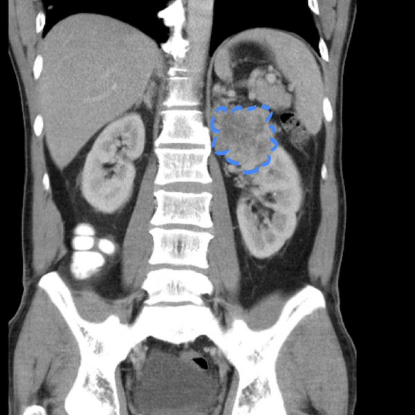
File:Appendicitis and renal cell carcinoma (Radiopaedia 17063-18402 C 1).png
Jump to navigation
Jump to search

Size of this preview: 600 × 600 pixels. Other resolutions: 240 × 240 pixels | 480 × 480 pixels | 699 × 699 pixels.
Original file (699 × 699 pixels, file size: 320 KB, MIME type: image/png)
Summary:
| Description |
|
| Date | Published: 13th Mar 2012 |
| Source | https://radiopaedia.org/cases/appendicitis-and-renal-cell-carcinoma |
| Author | Andrew Dixon |
| Permission (Permission-reusing-text) |
http://creativecommons.org/licenses/by-nc-sa/3.0/ |
Licensing:
Attribution-NonCommercial-ShareAlike 3.0 Unported (CC BY-NC-SA 3.0)
File history
Click on a date/time to view the file as it appeared at that time.
| Date/Time | Thumbnail | Dimensions | User | Comment | |
|---|---|---|---|---|---|
| current | 14:25, 20 May 2021 |  | 699 × 699 (320 KB) | Fæ (talk | contribs) | Radiopaedia project rID:17063 (batch #2691-3 C1) |
You cannot overwrite this file.
File usage
There are no pages that use this file.