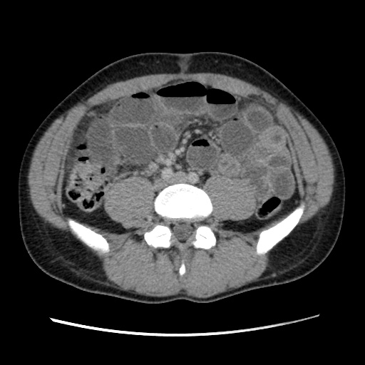
File:Appendicitis complicated by post-operative collection (Radiopaedia 35595-37114 A 53).jpg
Jump to navigation
Jump to search
Appendicitis_complicated_by_post-operative_collection_(Radiopaedia_35595-37114_A_53).jpg (512 × 512 pixels, file size: 62 KB, MIME type: image/jpeg)
Summary:
| Description |
|
| Date | Published: 17th Apr 2015 |
| Source | https://radiopaedia.org/cases/appendicitis-complicated-by-post-operative-collection |
| Author | Andrew Dixon |
| Permission (Permission-reusing-text) |
http://creativecommons.org/licenses/by-nc-sa/3.0/ |
Licensing:
Attribution-NonCommercial-ShareAlike 3.0 Unported (CC BY-NC-SA 3.0)
File history
Click on a date/time to view the file as it appeared at that time.
| Date/Time | Thumbnail | Dimensions | User | Comment | |
|---|---|---|---|---|---|
| current | 15:07, 20 May 2021 |  | 512 × 512 (62 KB) | Fæ (talk | contribs) | Radiopaedia project rID:35595 (batch #2692-53 A53) |
You cannot overwrite this file.
File usage
The following page uses this file:
