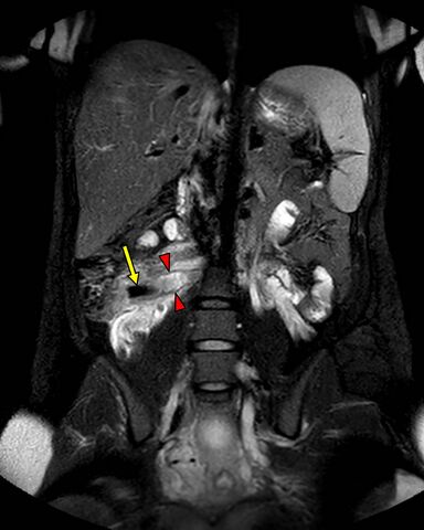
File:Appendicitis in gravida (MRI) (Radiopaedia 89433-106400 Coronal T2 SPAIR 1).jpg
Jump to navigation
Jump to search

Size of this preview: 480 × 600 pixels. Other resolutions: 192 × 240 pixels | 384 × 480 pixels | 615 × 768 pixels | 820 × 1,024 pixels | 1,456 × 1,819 pixels.
Original file (1,456 × 1,819 pixels, file size: 150 KB, MIME type: image/jpeg)
Summary:
| Description |
|
| Date | Published: 9th May 2021 |
| Source | https://radiopaedia.org/cases/appendicitis-in-gravida-mri |
| Author | Yair Glick |
| Permission (Permission-reusing-text) |
http://creativecommons.org/licenses/by-nc-sa/3.0/ |
Licensing:
Attribution-NonCommercial-ShareAlike 3.0 Unported (CC BY-NC-SA 3.0)
File history
Click on a date/time to view the file as it appeared at that time.
| Date/Time | Thumbnail | Dimensions | User | Comment | |
|---|---|---|---|---|---|
| current | 18:38, 20 May 2021 | | 1,456 × 1,819 (150 KB) | Fæ (talk | contribs) | Radiopaedia project rID:89433 (batch #2695-2 B1) |
You cannot overwrite this file.
File usage
There are no pages that use this file.
_(Radiopaedia_89433-106400_Coronal_T2_SPAIR_1).jpg&oldid=378230)