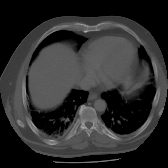
File:Appendicitis with perforation (Radiopaedia 35790-37342 Axial bone window 3).jpg
Jump to navigation
Jump to search
Size of this preview: 600 × 600 pixels. Other resolutions: 240 × 240 pixels | 480 × 480 pixels | 688 × 688 pixels.
Original file (688 × 688 pixels, file size: 63 KB, MIME type: image/jpeg)
Summary:
| Description |
|
| Date | Published: 26th Apr 2015 |
| Source | https://radiopaedia.org/cases/appendicitis-with-perforation-1 |
| Author | Andrew Dixon |
| Permission (Permission-reusing-text) |
http://creativecommons.org/licenses/by-nc-sa/3.0/ |
Licensing:
Attribution-NonCommercial-ShareAlike 3.0 Unported (CC BY-NC-SA 3.0)
File history
Click on a date/time to view the file as it appeared at that time.
| Date/Time | Thumbnail | Dimensions | User | Comment | |
|---|---|---|---|---|---|
| current | 21:38, 20 May 2021 | | 688 × 688 (63 KB) | Fæ (talk | contribs) | Radiopaedia project rID:35790 (batch #2709-55 B3) |
You cannot overwrite this file.
File usage
The following page uses this file:
.jpg&oldid=379355)