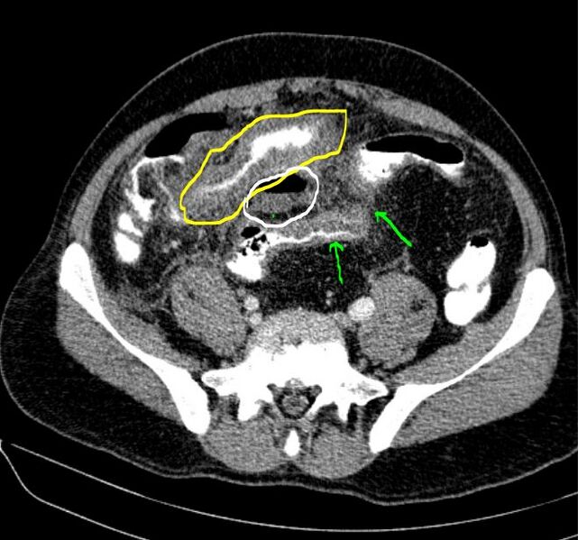
File:Appendicular phlegmon (Radiopaedia 80777-94269 axial CT scan 1).JPG
Jump to navigation
Jump to search

Size of this preview: 641 × 599 pixels. Other resolutions: 257 × 240 pixels | 514 × 480 pixels | 659 × 616 pixels.
Original file (659 × 616 pixels, file size: 78 KB, MIME type: image/jpeg)
Summary:
| Description |
|
| Date | Published: 4th Aug 2020 |
| Source | https://radiopaedia.org/cases/appendicular-phlegmon |
| Author | Faeze Salahshour |
| Permission (Permission-reusing-text) |
http://creativecommons.org/licenses/by-nc-sa/3.0/ |
Licensing:
Attribution-NonCommercial-ShareAlike 3.0 Unported (CC BY-NC-SA 3.0)
File history
Click on a date/time to view the file as it appeared at that time.
| Date/Time | Thumbnail | Dimensions | User | Comment | |
|---|---|---|---|---|---|
| current | 17:50, 21 May 2021 |  | 659 × 616 (78 KB) | Fæ (talk | contribs) | Radiopaedia project rID:80777 (batch #2733-1 A1) |
You cannot overwrite this file.
File usage
There are no pages that use this file.