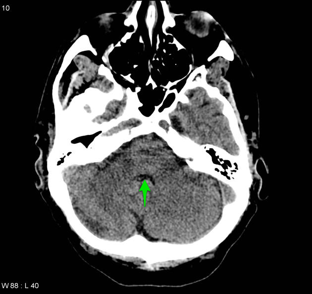
File:Aqueduct stenosis (Radiopaedia 5050-19529 D 1).png
Jump to navigation
Jump to search
Aqueduct_stenosis_(Radiopaedia_5050-19529_D_1).png (630 × 595 pixels, file size: 217 KB, MIME type: image/png)
Summary:
| Description |
|
| Date | Published: 25th Nov 2008 |
| Source | https://radiopaedia.org/cases/aqueduct-stenosis-5 |
| Author | Frank Gaillard |
| Permission (Permission-reusing-text) |
http://creativecommons.org/licenses/by-nc-sa/3.0/ |
Licensing:
Attribution-NonCommercial-ShareAlike 3.0 Unported (CC BY-NC-SA 3.0)
File history
Click on a date/time to view the file as it appeared at that time.
| Date/Time | Thumbnail | Dimensions | User | Comment | |
|---|---|---|---|---|---|
| current | 14:15, 22 May 2021 |  | 630 × 595 (217 KB) | Fæ (talk | contribs) | Radiopaedia project rID:5050 (batch #2771-4 D1) |
You cannot overwrite this file.
File usage
There are no pages that use this file.
