File:Aqueduct stenosis (Radiopaedia 52453-58460 Coronal T1 41).jpg

From NC Commons
Jump to navigation Jump to search

Aqueduct_stenosis_(Radiopaedia_52453-58460_Coronal_T1_41).jpg(512 × 512 pixels, file size: 42 KB, MIME type: image/jpeg)

Summary:

Description
  • Radiopaedia case ID: 52453
  • Image ID: 29687628
  • Image stack position: 41/58
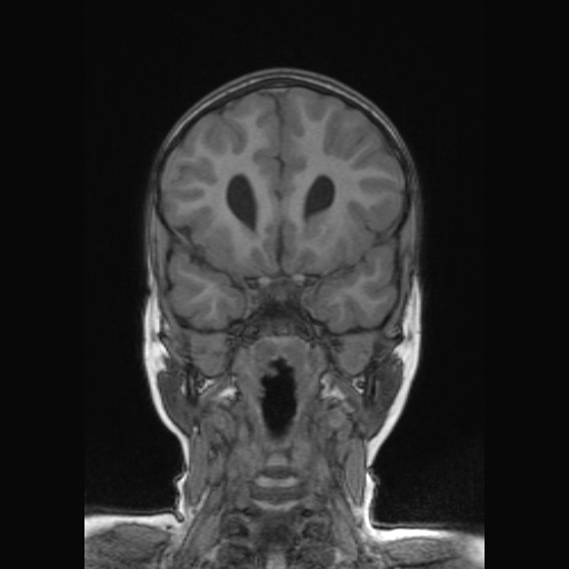
  • Plane projection: Coronal
  • Aux modality: T1
  • Study description: Post-natal MRI aged 1 year 4 months
  • Study findings: Typical features of aqueductal stenosis on sagittal T1 and Axial T2 weghted imaging, with dilatation of the lateral and now third ventricles.  There is thinning of the corpus callosum, especially posteriorly,  due to long term pressure effects. Note fornices have “fallen down” due to lack of support from the perforated septum pellucidum. Sulcal development is appropriate.
  • Modality: MRI
  • System: Obstetrics
  • Findings: Selected ultrasound images at 21 weeks gestation demonstrating bilateral ventriculomegaly, measuring 16 and 18mm. Cavum septum pellucidum is not identified. Complete study was not available. The cerebellum and posterior fossa were reported normal. The third ventricle appeared prominent, measuring 2.5 mm.  Detailed survey of the rest of the fetal anatomy showed no additional abnormality. Female fetal gender.
Date Published: 12th Apr 2017
Source https://radiopaedia.org/cases/aqueduct-stenosis
Author Alexandra Stanislavsky
Permission
(Permission-reusing-text)
http://creativecommons.org/licenses/by-nc-sa/3.0/

Licensing:

Attribution-NonCommercial-ShareAlike 3.0 Unported (CC BY-NC-SA 3.0)

File history

Click on a date/time to view the file as it appeared at that time.

Date/TimeThumbnailDimensionsUserComment
current12:10, 22 May 2021Thumbnail for version as of 12:10, 22 May 2021512 × 512 (42 KB) (talk | contribs)Radiopaedia project rID:52453 (batch #2769-140 C41)

The following page uses this file: