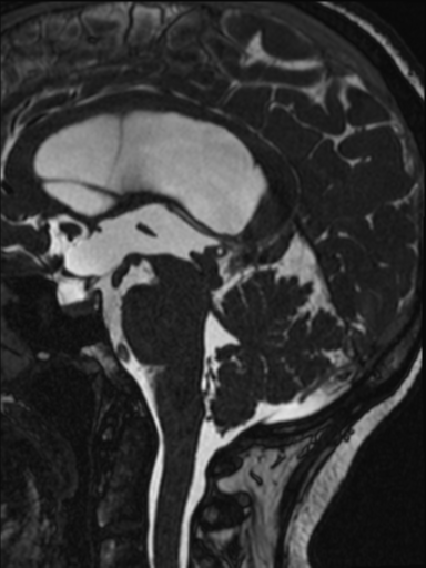
File:Aqueduct stenosis - web (Radiopaedia 59840-67276 Sagittal T2 35).png
Jump to navigation
Jump to search
Aqueduct_stenosis_-_web_(Radiopaedia_59840-67276_Sagittal_T2_35).png (384 × 512 pixels, file size: 88 KB, MIME type: image/png)
Summary:
| Description |
|
| Date | Published: 17th May 2018 |
| Source | https://radiopaedia.org/cases/aqueduct-stenosis-web-1 |
| Author | Frank Gaillard |
| Permission (Permission-reusing-text) |
http://creativecommons.org/licenses/by-nc-sa/3.0/ |
Licensing:
Attribution-NonCommercial-ShareAlike 3.0 Unported (CC BY-NC-SA 3.0)
File history
Click on a date/time to view the file as it appeared at that time.
| Date/Time | Thumbnail | Dimensions | User | Comment | |
|---|---|---|---|---|---|
| current | 17:58, 22 May 2021 |  | 384 × 512 (88 KB) | Fæ (talk | contribs) | Radiopaedia project rID:59840 (batch #2778-35 A35) |
You cannot overwrite this file.
File usage
The following page uses this file:
