File:Aqueductal stenosis (Radiopaedia 15736).jpg
Jump to navigation
Jump to search
Size of this preview: 600 × 600 pixels. Other resolutions: 240 × 240 pixels | 480 × 480 pixels.
Original file (800 × 800 pixels, file size: 229 KB, MIME type: image/jpeg)
Summary:
- Radiopaedia case ID: 15736
- Image ID: 1470958
- Study description: MRI brain - single image
- Modality: MRI
- System: Central Nervous System
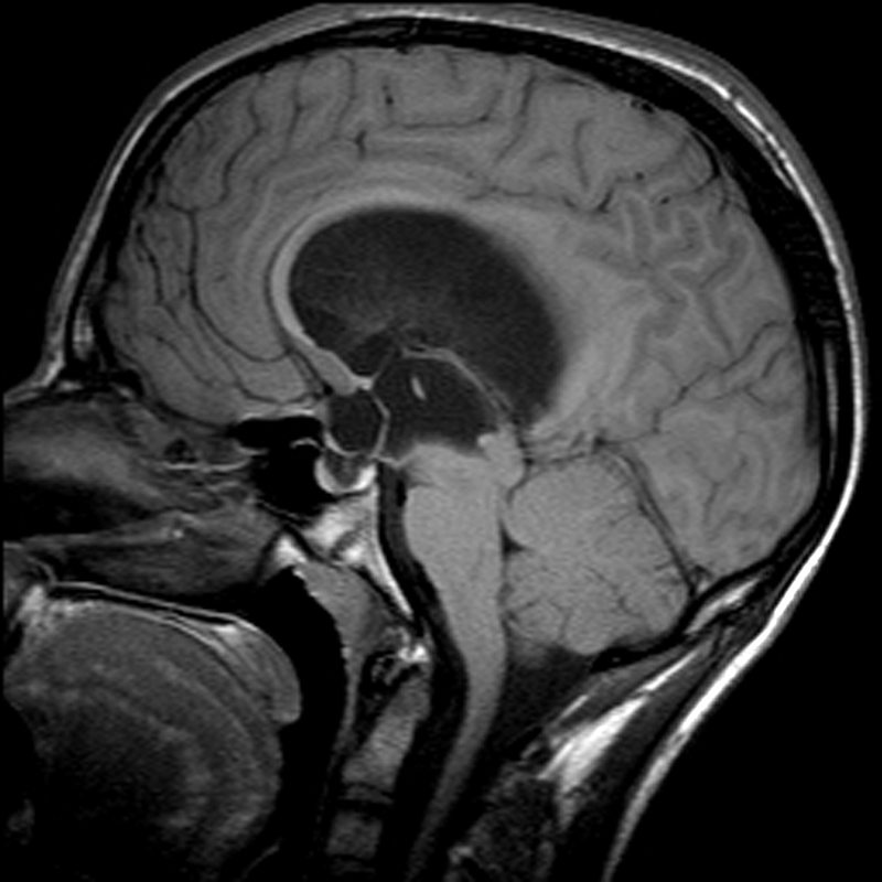
- Findings: Midline sagittal T1 weighted image demonstrates marked hydrocephalus, with upward bowing and thinning of the corpus callosum, fenestration of the septum pellucidum and ballooning out of the third ventricular recesses. The superior part of the aqueduct is funnelled, whereas the distal part is stenosed.
- Published: 5th Nov 2011
- Source: https://radiopaedia.org/cases/aqueductal-stenosis-3
- Author: Frank Gaillard
- Permission: http://creativecommons.org/licenses/by-nc-sa/3.0/
Licensing:
Attribution-NonCommercial-ShareAlike 3.0 Unported (CC BY-NC-SA 3.0)
File history
Click on a date/time to view the file as it appeared at that time.
| Date/Time | Thumbnail | Dimensions | User | Comment | |
|---|---|---|---|---|---|
| current | 23:24, 18 March 2021 | | 800 × 800 (229 KB) | Fæ (talk | contribs) | Radiopaedia project rID:15736 (batch #2703) |
You cannot overwrite this file.
File usage
There are no pages that use this file.
.jpg&oldid=8859797)