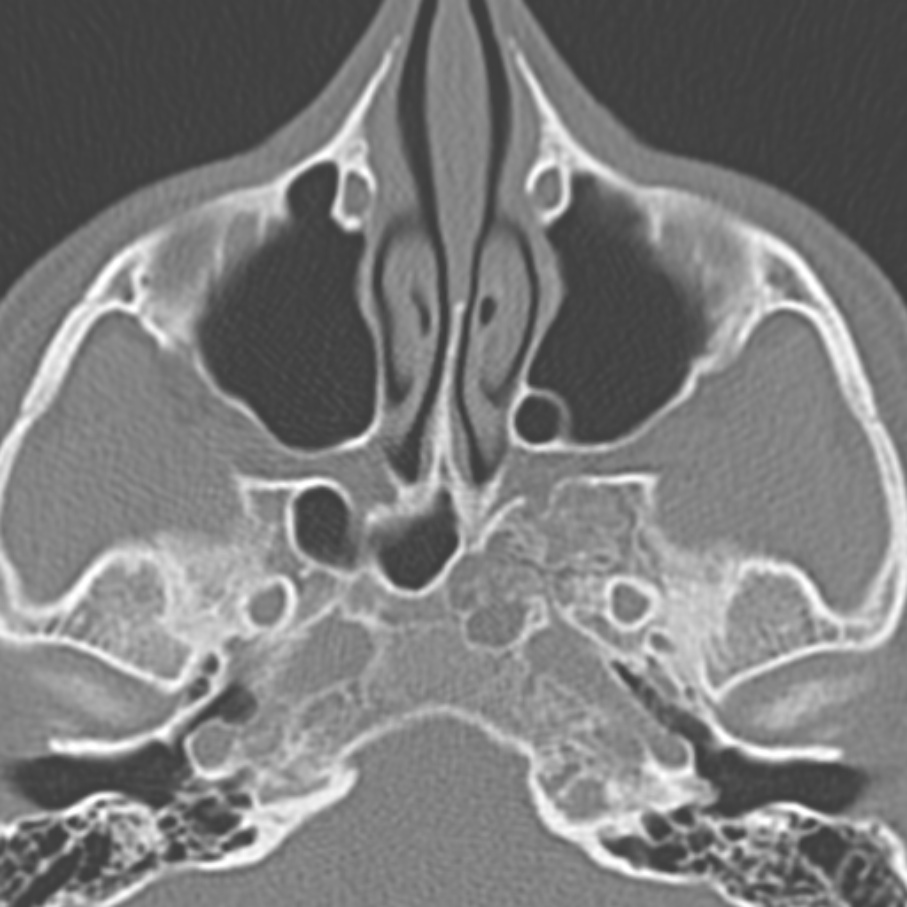
File:Arrested pneumatization of the sphenoid (Radiopaedia 10700-11172 Axial non-contrast 7).jpg
Jump to navigation
Jump to search
Size of this preview: 600 × 600 pixels. Other resolutions: 240 × 240 pixels | 480 × 480 pixels | 907 × 907 pixels.
Original file (907 × 907 pixels, file size: 154 KB, MIME type: image/jpeg)
Summary:
| Description |
|
| Date | Published: 7th Sep 2010 |
| Source | https://radiopaedia.org/cases/arrested-pneumatisation-of-the-sphenoid-2 |
| Author | Erik Ranschaert |
| Permission (Permission-reusing-text) |
http://creativecommons.org/licenses/by-nc-sa/3.0/ |
Licensing:
Attribution-NonCommercial-ShareAlike 3.0 Unported (CC BY-NC-SA 3.0)
File history
Click on a date/time to view the file as it appeared at that time.
| Date/Time | Thumbnail | Dimensions | User | Comment | |
|---|---|---|---|---|---|
| current | 05:09, 24 May 2021 | | 907 × 907 (154 KB) | Fæ (talk | contribs) | Radiopaedia project rID:10700 (batch #2903-7 A7) |
You cannot overwrite this file.
File usage
There are no pages that use this file.
.jpg&oldid=401256)