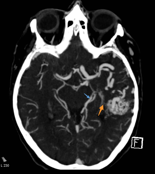
File:Arteriovenous malformation - cerebral (Radiopaedia 8172-14683 Figure 3 1).png
Jump to navigation
Jump to search
Arteriovenous_malformation_-_cerebral_(Radiopaedia_8172-14683_Figure_3_1).png (535 × 600 pixels, file size: 208 KB, MIME type: image/png)
Summary:
| Description |
|
| Date | Published: 17th Jan 2010 |
| Source | https://radiopaedia.org/cases/arteriovenous-malformation-cerebral-1 |
| Author | Frank Gaillard |
| Permission (Permission-reusing-text) |
http://creativecommons.org/licenses/by-nc-sa/3.0/ |
Licensing:
Attribution-NonCommercial-ShareAlike 3.0 Unported (CC BY-NC-SA 3.0)
File history
Click on a date/time to view the file as it appeared at that time.
| Date/Time | Thumbnail | Dimensions | User | Comment | |
|---|---|---|---|---|---|
| current | 01:13, 25 May 2021 |  | 535 × 600 (208 KB) | Fæ (talk | contribs) | Radiopaedia project rID:8172 (batch #2938-3 C1) |
You cannot overwrite this file.
File usage
There are no pages that use this file.
