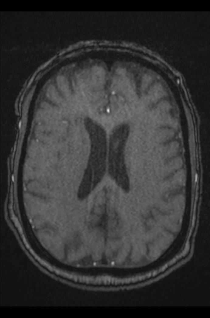
File:Artery of Percheron infarction (Radiopaedia 80613-94409 Axial MRA 118).jpg

From NC Commons
Jump to navigation Jump to search

Original file(691 × 1,047 pixels, file size: 44 KB, MIME type: image/jpeg)

Summary:

Description
  • Radiopaedia case ID: 80613
  • Image ID: 53263356
  • Image stack position: 118/143
  • Plane projection: Axial
  • Aux modality: MRA
  • Study findings: Areas of diffusion restriction noted in biparamedian ventro-medial thalami and in left paramedian rostral midbrain showing signal dropout on ADC images. These areas appear faintly high on T2 and FLAIR images and iso to hypo-intense on T1W images. Imaging appearances are consistent with artery of percheron infarct. Mild to moderate micro-vascular ischemic demyelinations and few old lacunar infarcts noted in white matter bilaterally. Age related involutional changes seen in brain with deepened cortical sulci, slightly widened extra-axial CSF spaces and mild prominence of ventricular system. Faint high intensity signal return noted from medullaoblongata only appreciated on FLAIR coronal images is likely be artifactual as it is not appreciated on rest of the sequences. Hypertrophied nasal turbinates and mildly deviated median nasal septum noted to the left side. Poor pneumatization of mastoid air cells noted bilaterally. MRA TOF Mild to moderate atherosclerotic disease seen in M2 and M3 segments of left middle cerebral artery and mild in few of peripheral cortical branches of both middle cerebral arteries. A1 segment of left anterior cerebral artery appears hypoplastic. Reduced caliber of A2 segment of left anterior cerebral artery also noted may be hypoplastic or secondary to atherosclerotic disease. Reduced caliber and intermittent paucity of signal return noted from right vertebral artery likely be hypoplastic with mild associated atherosclerotic disease. Mild short segment atherosclerotic disease also seen in basilar artery and in P1 segment of left posterior cerebral artery. Rest of the MRI study is normal.
  • Modality: MRI
  • System: Vascular
  • Findings: Few lacunar infarcts are seen in white matter of both frontal lobes, left thalamus and mid brain. Mild microvascular ischemic changes are seen in white matter bilaterally. Age appropriate involutional changes are seen with deepened cortical sulci and prominence of sylvian fissures. Normal both orbits. Mild mucosal thickening is noted in posterior ethmoid air cells. Hypertrophied right inferior nasal turbinates. Deviated median nasal septum convex to left side. Rest of the scan is normal.
Date Published: 20th Aug 2020
Source https://radiopaedia.org/cases/artery-of-percheron-infarction-10
Author Muhammad Qasim Naeem
Permission
(Permission-reusing-text)
http://creativecommons.org/licenses/by-nc-sa/3.0/

Licensing:

Attribution-NonCommercial-ShareAlike 3.0 Unported (CC BY-NC-SA 3.0)

File history

Click on a date/time to view the file as it appeared at that time.

Date/TimeThumbnailDimensionsUserComment
current08:29, 25 May 2021Thumbnail for version as of 08:29, 25 May 2021691 × 1,047 (44 KB) (talk | contribs)Radiopaedia project rID:80613 (batch #2958-361 K118)