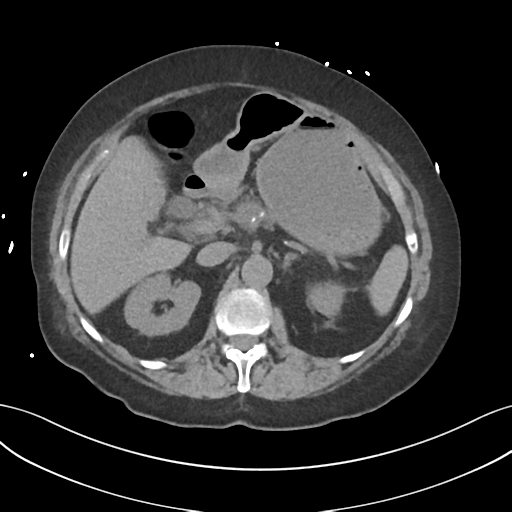
File:Ascending cholangitis (Radiopaedia 39068-41253 Axial non-contrast 18).png
Jump to navigation
Jump to search
Ascending_cholangitis_(Radiopaedia_39068-41253_Axial_non-contrast_18).png (512 × 512 pixels, file size: 165 KB, MIME type: image/png)
Summary:
| Description |
|
| Date | Published: 18th Aug 2015 |
| Source | https://radiopaedia.org/cases/ascending-cholangitis |
| Author | Henry Knipe |
| Permission (Permission-reusing-text) |
http://creativecommons.org/licenses/by-nc-sa/3.0/ |
Licensing:
Attribution-NonCommercial-ShareAlike 3.0 Unported (CC BY-NC-SA 3.0)
File history
Click on a date/time to view the file as it appeared at that time.
| Date/Time | Thumbnail | Dimensions | User | Comment | |
|---|---|---|---|---|---|
| current | 16:01, 26 May 2021 |  | 512 × 512 (165 KB) | Fæ (talk | contribs) | Radiopaedia project rID:39068 (batch #3019-18 A18) |
You cannot overwrite this file.
File usage
The following page uses this file:
