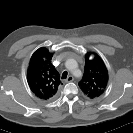
File:Aspirated tooth (Radiopaedia 28584-28844 Axial C+ CTPA 20).jpg

From NC Commons
Jump to navigation Jump to search

Aspirated_tooth_(Radiopaedia_28584-28844_Axial_C+_CTPA_20).jpg(512 × 512 pixels, file size: 28 KB, MIME type: image/jpeg)

Summary:

Description
  • Radiopaedia case ID: 28584
  • Image ID: 6531892
  • Image stack position: 20/72
  • Plane projection: Axial
  • Aux modality: C+ CTPA
  • Study description: CTPA was requested by MET call team prior to reviewing chest radiograph
  • Study findings: A radiodense structure consistent with a tooth is lodged in the left lower lobe bronchus. No overt collapse is present at this time. The tooth is present on radiograph this morning but was not present on yesterdays CXR. When compared to prior imaging, there is mildly progressive air space consolidation in both lower lobes, and there are progressive bilateral pleural effusions with tracking upward between the fissures. Segmental pulmonary emboli seen in all right lobe segments apart from the posterior and lateral ones. Superior lingular segmantal emboli on the left. Serial left- sided non-displaced rib fracture. No pneumothorax. Conclusion: Left lower lobe bronchus foreign body consistent with a tooth in this patient with dental trauma. Multiple PEs as described.
  • Modality: CT
  • System: Chest
  • Findings: No acute abnormality.
Date Published: 6th Apr 2014
Source https://radiopaedia.org/cases/aspirated-tooth-1
Author Jan Frank Gerstenmaier
Permission
(Permission-reusing-text)
http://creativecommons.org/licenses/by-nc-sa/3.0/

Licensing:

Attribution-NonCommercial-ShareAlike 3.0 Unported (CC BY-NC-SA 3.0)

File history

Click on a date/time to view the file as it appeared at that time.

Date/TimeThumbnailDimensionsUserComment
current05:39, 27 May 2021Thumbnail for version as of 05:39, 27 May 2021512 × 512 (28 KB) (talk | contribs)Radiopaedia project rID:28584 (batch #3086-21 B20)

The following page uses this file: