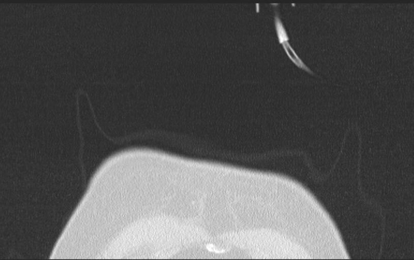
File:Aspirated tooth (Radiopaedia 28584-28844 Axial lung window 13).jpg
Jump to navigation
Jump to search
Size of this preview: 800 × 503 pixels. Other resolutions: 320 × 201 pixels | 640 × 403 pixels | 814 × 512 pixels.
Original file (814 × 512 pixels, file size: 41 KB, MIME type: image/jpeg)
Summary:
| Description |
|
| Date | Published: 6th Apr 2014 |
| Source | https://radiopaedia.org/cases/aspirated-tooth-1 |
| Author | Jan Frank Gerstenmaier |
| Permission (Permission-reusing-text) |
http://creativecommons.org/licenses/by-nc-sa/3.0/ |
Licensing:
Attribution-NonCommercial-ShareAlike 3.0 Unported (CC BY-NC-SA 3.0)
File history
Click on a date/time to view the file as it appeared at that time.
| Date/Time | Thumbnail | Dimensions | User | Comment | |
|---|---|---|---|---|---|
| current | 06:00, 27 May 2021 | | 814 × 512 (41 KB) | Fæ (talk | contribs) | Radiopaedia project rID:28584 (batch #3086-167 D13) |
You cannot overwrite this file.
File usage
The following page uses this file:
.jpg&oldid=422435)