File:Aspirated tooth (Radiopaedia 73513).jpg
Jump to navigation
Jump to search

Size of this preview: 339 × 600 pixels. Other resolutions: 135 × 240 pixels | 499 × 883 pixels.
Original file (499 × 883 pixels, file size: 69 KB, MIME type: image/jpeg)
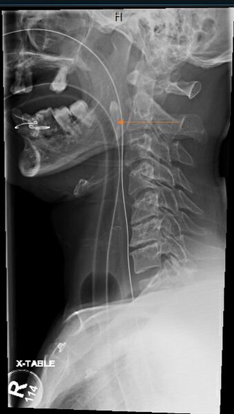
Summary:
- Radiopaedia case ID: 73513
- Image ID: 51919231
- Study findings: Lateral neck x-ray: Partially visualized proximal aspect of nasogastric and endotracheal tubes. There is a shadow of a tooth seen in the region of oropharynx between both tubes at the level of C2 (arrow). Multiple loose upper jaw teeth with defect between the teeth noted. Multilevel degenerative changes of the cervical spine.
- Modality: X-ray
- System: Trauma
- Findings: Lateral neck x-ray: Partially visualized proximal aspect of nasogastric and endotracheal tubes. There is a shadow of a tooth seen in the region of oropharynx between both tubes at the level of C2 (arrow). Multiple loose upper jaw teeth with defect between the teeth noted. Multilevel degenerative changes of the cervical spine.
- Published: 13th Jan 2020
- Source: https://radiopaedia.org/cases/aspirated-tooth-4
- Author: Ghufran Aref Saeed
- Permission: http://creativecommons.org/licenses/by-nc-sa/3.0/
Licensing:
Attribution-NonCommercial-ShareAlike 3.0 Unported (CC BY-NC-SA 3.0)
File history
Click on a date/time to view the file as it appeared at that time.
| Date/Time | Thumbnail | Dimensions | User | Comment | |
|---|---|---|---|---|---|
| current | 09:56, 19 March 2021 | | 499 × 883 (69 KB) | Fæ (talk | contribs) | Radiopaedia project rID:73513 (batch #3014) |
You cannot overwrite this file.
File usage
There are no pages that use this file.
.jpg&oldid=8859766)