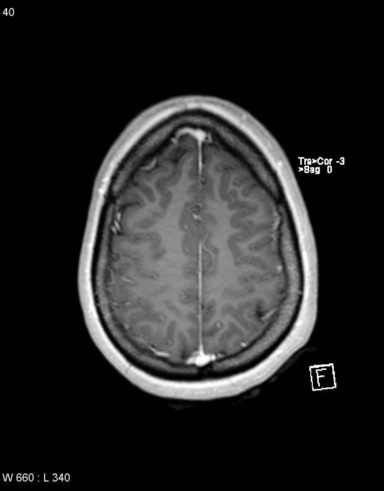
File:Astroblastoma (Radiopaedia 39792-42218 Axial T1 C+ 39).jpg
Jump to navigation
Jump to search
Size of this preview: 468 × 599 pixels. Other resolutions: 187 × 240 pixels | 375 × 480 pixels | 766 × 981 pixels.
Original file (766 × 981 pixels, file size: 55 KB, MIME type: image/jpeg)
Summary:
| Description |
|
| Date | Published: 21st Sep 2015 |
| Source | https://radiopaedia.org/cases/astroblastoma |
| Author | Alan Coulthard |
| Permission (Permission-reusing-text) |
http://creativecommons.org/licenses/by-nc-sa/3.0/ |
Licensing:
Attribution-NonCommercial-ShareAlike 3.0 Unported (CC BY-NC-SA 3.0)
File history
Click on a date/time to view the file as it appeared at that time.
| Date/Time | Thumbnail | Dimensions | User | Comment | |
|---|---|---|---|---|---|
| current | 22:06, 27 May 2021 | | 766 × 981 (55 KB) | Fæ (talk | contribs) | Radiopaedia project rID:39792 (batch #3104-39 A39) |
You cannot overwrite this file.
File usage
The following page uses this file:
.jpg&oldid=424907)